Rabbit polyclonal antibody to neurofilament NF-H
Rat embyro cerebral cultures stained with RPCA-NF-H-Ind, our phosphate independent rabbit polyclonal antibodies to neurofilamcent NF-H (green), and counterstained with our chicken antibody to GFAP (CPCA-GFAP, red). The NF-H antibody stains is phosphate independent and so recognizes axonal, dendritic and perikaryal neurofilaments, so the full morphology of neurons can be seen. The GFAP antibody stains surrounding astrocytic cells. Nuclei are stained with a DNA intercalating agent (blue). Link here back to the EnCor Biotechnology Home Page.
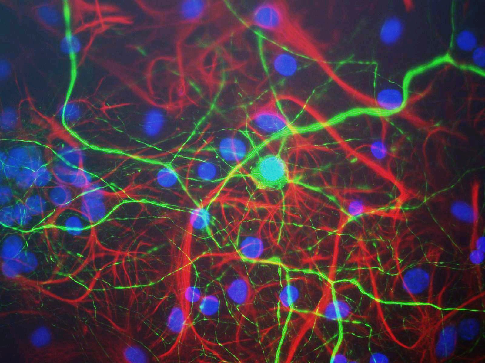
The RPCA-NF-H-Pind antibody was used at a dilution of 1:1,000, as was the CPCA-GFAP antibody. The secondary antibodies were from Molecular Probes/Invitrogen. Cultures were processed using our standard fixation and staining procedure (described here). To order these antibodies go to our order form (here). Picture taken with a Zeiss 40X objective and documented with a Diagnostic Instrument RT-Slider camera.
©EnCor Biotechnology Inc. .